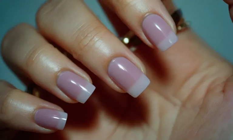
Can You Cut Off A Mole With Nail Clippers? A Detailed Guide
Moles are extremely common, with most adults having between 10 to 40 moles. While moles are usually harmless, some changes like growth, color changes or itching can indicate skin cancer. This often leads people to want to remove moles at home.
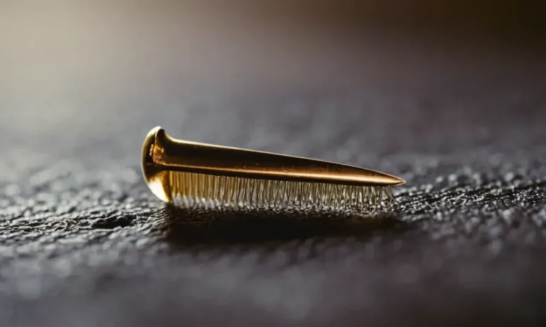
If you’re wondering if using nail clippers is an option, here’s a quick answer: cutting off a mole with nail clippers is never recommended and can be dangerous. Instead, it’s best to have a mole professionally removed by a dermatologist.
In this comprehensive guide, we’ll cover everything you need to know about safely removing moles. We’ll discuss the risks of using nail clippers, when moles should be removed, the proper mole removal techniques used by doctors, aftercare instructions and more.
Why You Should Not Use Nail Clippers on Moles
Can Lead to Infection and Scarring
Using nail clippers on a mole can potentially introduce bacteria and trigger an infection. The serrated edges of nail clippers create an open wound that is vulnerable to bacteria growth. According to the CDC, only sterilized medical instruments should come in contact with open skin to prevent contamination.
An infected mole may scab over, ooze pus, become tender and swollen – ultimately resulting in an unattractive scar if not properly treated with antibiotics.
Does Not Remove the Entire Mole
Nail clippers only snip off the visible part of a mole above the skin’s surface. However, the root and cell clusters that make up the mole extend much deeper into the dermis layer. Any remnants left under the skin will likely continue growing over time.
For complete extraction, a biopsy is required along with stitches to close the wound. Using nail clippers leads to a mole growing back – usually larger and darker than before.
High Risk of Bleeding
Digging into a mole with nail clippers often causes profuse bleeding that is difficult to stop at home. The abundance of blood vessels supplying nutrients to the mole means a laceration can result in continuous oozing of blood.
Applying pressure may temporarily suppress bleeding, but caution is needed as too much pressure can damage surrounding tissue. Uncontrolled blood loss warrants an emergency hospital visit for suturing and blood transfusion in extreme cases.
Painful With No Anesthetic
Attempting to cut or tear skin tissue with nail clippers is an intensely painful experience as the clipper blades slice through nerve endings. There is generally no numbing agent applied to diminish discomfort. The face and neck areas often chosen for mole removal have heightened sensitivities.
Patients undergoing professional mole removal are given local anesthetic injections around the site for pain relief during and after the procedure.
Does Not Allow for Pathology Testing
Chopping off a suspicious looking mole destroys tissue integrity making it impossible to have pathology analysis done after removal. Testing by a dermatopathologist is crucial for accurately diagnosing if skin cells are cancerous or benign.
Early melanoma detection and stages can only be confirmed through microscopic examination of the extracted mole. Using nail clippers eliminates this path for gaining insights about one’s skin health and any underlying conditions.
When to Get a Mole Removed
Change in Appearance, Color, Size
One of the most common reasons to get a mole removed is if it changes appearance. This includes changing color from brown to black or blue, getting larger or smaller, or changing shape. If you notice any new or unusual changes in an existing mole, it’s important to see your dermatologist right away as this could indicate skin cancer.
Your dermatologist can examine the mole and determine if a biopsy or removal is recommended.
According to the American Academy of Dermatology, moles that are bigger than the size of a pencil eraser, have irregular borders, contain different colors, or just look unusual are more likely to become cancerous.
It’s best not to ignore changes in your moles, as catching melanoma skin cancer early leads to a good prognosis.
New Symptoms Like Itching or Bleeding
In addition to physical changes, new symptoms can also indicate it’s time to see a dermatologist about mole removal. If a mole starts itching, hurting, oozing, or bleeding, make an appointment right away.
These symptoms can occur as a mole progresses to melanoma, so it’s important to get checked out sooner rather than later.
According to the Skin Cancer Foundation, itching is actually one of the most common symptoms of melanoma. Bleeding moles can also signal cancerous changes. Don’t ignore new sensations or symptoms associated with your moles.
For Cosmetic Reasons
Moles don’t need to be cancerous or bothersome to warrant removal. Many people elect to have benign moles taken off simply for cosmetic reasons. Moles that are large, raised, or in prominent locations like the face are often removed for aesthetic purposes.
In one survey published in 2021, nearly one-third of dermatologic surgery patients cited improved appearance as the reason for getting a mole removed. For many, mole removal can boost self-confidence. Just make sure to see a board-certified dermatologist to ensure it is done safely.
Mole Looks Abnormal or Irregular
Finally, getting a mole removed as a precaution due to an unusual appearance is sensible. Even if a mole isn’t changing or symptomatic, an abnormal shape, border, color, or diameter can indicate precancerous or cancerous changes.
Dermatologists recommend the ABCDE method to assess if a mole looks irregular:
- Asymmetry – One half is unlike the other half
- Border – The edges are ragged or blurred
- Color – There is more than one color present
- Diameter – It is larger than 6mm, about the size of a pencil eraser
- Evolving – Any change in size, shape, symptoms
If a mole exhibits any of these traits, it is considered atypical and removal is usually recommended. It is always better to be safe and have an unusual mole evaluated and taken off when necessary.
Professional Mole Removal Techniques
Excisional Surgery
Excisional surgery is considered the gold standard for mole removal. This technique involves numbing the area around the mole and then using a scalpel to cut out the entire mole along with a margin of normal-looking skin around it. The skin is then closed with stitches.
Excisional surgery has the highest cure rate for suspicious moles and allows the entire mole to be examined under a microscope to check margins and determine if the mole was cancerous. Excisional surgery does leave a scar, so it may not be the best option for moles in cosmetically sensitive areas.
Punch Biopsy
A punch biopsy uses a special surgical tool that looks like a tiny cookie cutter to remove a circular core of skin that contains the mole. After numbing the skin, the doctor uses the punch biopsy tool to cut down through the skin and remove a column of tissue with the mole at the bottom.
This leaves an open hole in the skin that is closed with stitches. A punch biopsy removes the entire thickness of the mole so it can be sent for microscopic analysis, but it does not remove any extra margin around the mole. So if cancer cells are seen near the edges, more surgery may be needed.
Because it takes out less tissue, a punch biopsy causes a little less scarring than excisional surgery.
Shave Removal
Shave removal, also called shave biopsy or shave excision, is best for small moles that stand up above the surface of the skin. After numbing the skin, the doctor uses a sharp surgical blade to slice off the mole.
The removed tissue is very thin since it shaves off only the part sticking up above the skin surface. This means the mole cannot be fully examined under the microscope to check margins and determine if it is cancerous.
Shave removal is less invasive than other techniques so it usually leaves minimal scarring. But it may need to be repeated if some mole tissue is left behind or if the biopsy comes back positive for cancer cells.
Laser Removal
Laser mole removal uses focused laser light to vaporize moles. A laser targets melanin, the brown pigment in moles, and heats it up until the cells burst. This destroys the mole with minimal damage to surrounding skin. Laser removal requires no cutting or sutures.
It can be done quickly in a doctor’s office. Compared to surgical excision, laser removal results in less pain, bleeding, and scarring. But lasers cannot remove deep moles or provide tissue samples for biopsies. Most laser removals require 2-3 treatments spaced a few weeks apart.
Laser removal is not recommended for suspected skin cancers since it destroys tissue needed for microscopic examination.
Cryosurgery
Cryosurgery freezes moles with liquid nitrogen to destroy abnormal cells. The doctor swabs or sprays liquid nitrogen directly onto the mole and surrounding margin of skin. This causes the cells to crystalize and die. The dead tissue then blisters and crusts and eventually falls off.
Cryotherapy mole removal does not require anesthesia or leave stitches. However, it can cause depigmentation or even damage in deeper layers of healthy skin. It is not recommended for suspected melanomas. Follow-up treatments are usually needed to fully remove all mole tissue.
Cryosurgery may be preferred for benign flat moles in children or elderly patients since it is a simple procedure with minimal scarring.
Aftercare Instructions Post Mole Removal
Keep the Area Clean
Keeping the wound site clean after mole removal is crucial to prevent infection and ensure proper healing. Gently clean the area daily with mild soap and water, dabbing it dry with a clean towel. Avoid rubbing or scratching the sensitive skin.
After a week, you can stop regular cleaning unless drainage, redness or swelling occurs. Covering it with sterile gauze helps protect from irritants. Check for signs of complications and contact your dermatologist if any arise.
Use Antibiotic Ointment
Applying antibiotic ointment aids healing post-procedure. Polysporin or Bacitracin are common options. Gently spread a thin layer over the wound using a cotton swab, up to 3 times a day. This prevents bacterial infection and creates a protective barrier allowing new skin to form.
Continue use for 1-2 weeks based on your practitioner’s advice. Stop if rashes, itching or redness develop. Allergic reactions can occasionally occur with topical antibiotics. Inform your dermatologist promptly about any issues.
Apply Cold Compress to Reduce Swelling
It’s normal to experience some swelling, redness and bruising after mole removal. To minimize discomfort, use a cold compress or ice pack wrapped in cloth on the site for 10-15 minutes several times a day. The cold constricts blood vessels, reducing inflammation and pain signals.
Don’t apply ice directly as it can damage tender skin. Discomfort typically peaks in the first 48 hours post-procedure then gradually subsides. If severe or persistent, contact your healthcare provider.
Avoid Irritating the Area
Preventing trauma to the wound is vital for optimal healing after mole extraction. Wear loose-fitting clothing over the site and avoid restrictive bandages. Protect it from friction with bandaids or gauze when necessary. Don’t scratch, pick or peel scabs as this can cause scarring or infection.
Avoid exposing the area to direct sun, harsh soaps or chemicals until fully healed. Follow activity restrictions from your practitioner, often involving avoiding heavy lifting, yardwork and rigorous exercise initially. Take care not to bump or traumatize the sensitive skin.
Watch for Signs of Infection
Monitor for symptoms like increasing pain, redness, swelling, warmth or pus which may indicate a wound infection requiring prompt medical care. Other concerning signs include foul odor, blueish skin discoloration or spread of redness beyond the site. Fever can occur with a spreading skin infection.
Contact your dermatologist immediately if you observe any of these issues starting 1-2 weeks post-procedure. Oral antibiotics or surgical drainage may be necessary to treat infected mole removal sites. Catching problems early is key to prevent complications.
Conclusion
While it may be tempting to use nail clippers to remove a mole yourself, this can lead to significant complications like scarring, bleeding and infection. It’s critical to have any concerning or unwanted moles examined and removed properly by a dermatologist.
Following your dermatologist’s instructions for aftercare can help ensure your removal site heals quickly and safely. With the right mole removal and aftercare, you can have clear skin without the risks of at-home methods.
If you have any moles causing concern, schedule an appointment with your dermatologist. They can determine the best course of action for examination and safe removal.